Retinal degenerative diseases: critical phase in the differentiative pathway of photoreceptors identified
The research, published in Scientific Reports, is the result of a collaboration between the University of Pisa and the Sant'Anna School of Advanced Studies

Retinal photoreceptors, the cells in the eye that function as light sensors, develop their specific shapes and functions based on instructions received at the time of their generation from multipotent stem cells. However, a study conducted by researchers at the University of Pisa and the Sant'Anna School of Advanced Studies showed that if, during a limited time interval following their initial fate assignment, they do not receive specific signals of both physical and chemical types from the environment, then they will develop hybrid features between photoreceptors and glial cells. The results of the study were published in the international journal Scientific Reports in an article titled "Increasing cell culture density during a developmental window prevents fated rod precursors derailment toward hybrid rod-glia cells", with authors Massimiliano Andreazzoli (Department of Biology, UniPi), Gian Carlo Demontis (Department of Pharmacy, UniPi) and Debora Angeloni ( Sant'Anna School).
The study was aimed at understanding the reasons why, in preclinical models of replacement therapies for retinal degenerative diseases, most transplanted cells fail to integrate into the recipient's retina to effectively replace degenerated cells.
The study is part of a larger project coordinated by Vania Broccoli (CNR Neurophysiology Institute and San Raffaele) and funded by the Fondazione Roma. It started in 2016 and grew out of Andreazzoli's interest in molecular genetics mechanisms of retinal development, Demontis' interest in electrophysiological function of photoreceptors, and Angeloni's interest in cellular mechanotransduction mechanisms.
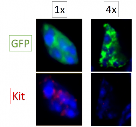
In gallery: 2 retinal rod precursors, recognizable by the expression of a green fluorescent protein (GFP), cultured at 2 different densities (cells per unit area - 1x and 4x). The expression of an immaturity gene (Kit - indicated by red color), persists in the precursor cultured at the lower density (1x), while it disappears in the precursor cultured at the higher density (4x). The cell nucleus is stained blue.


